No Insurance? No Problem! Join Our Advantage Plan
3D Cone Beam and Dental Scans
Advancements in dental technology have brought about many new tools that make it easier for dentists and more comfortable for patients. Dental cone beam computed tomography (CBCT), a new type of X-ray equipment, allows dentists to see a clear, detailed, three-dimensional image of the mouth without causing pain to the patient. This 3D imaging system takes full photos of the teeth, soft tissues, nerve pathways, and bones in a single scan.
Cone beam dental scans are available at Dental Care of Morrisville in Morrisville and the surrounding area. Our staff can help you learn more about the procedure you are undergoing and answer any questions you have about 3D imaging. Call us at (919) 588-3000 to schedule a consultation appointment today.
How 3D Imaging Works
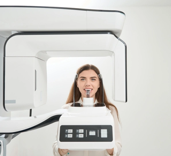
A 3D cone beam machine resembles conventional CT scan machines and comes in two different structures: an upright chair for sitting or a moveable table for lying down. Depending on the procedure and type of machine being used, the patient will be seated in an exam chair or lie down on an exam table. The chair has an extendable arm (C-arm) while the table has a rotator (gantry) that both rotate 360 degrees around the patient’s head, taking multiple images at once.
The images are taken at different angles and gathered to create a single 3D image. According to the Radiology organization, "In a single rotation, the detector can generate anywhere between 150 to 200 high resolution two-dimensional (2-D) images, which are then digitally combined to form a 3-D image that can provide your dentist or oral surgeon with valuable information about your oral and craniofacial health." The 3D image is available as soon as the scan is complete, allowing the doctor to discuss the patient’s treatment plan in the same visit.
“The chair has an extendable arm (C-arm) while the table has a rotator (gantry) that both rotate 360 degrees around the patient’s head, taking multiple images at once.”
Differences Between Traditional and Cone Beam
Traditional CT scanners and cone beam CT scanners both undertake the same basic function, but technical differences set them apart.
Traditional CT Scans
A traditional CT scan takes several pictures of internal body structures from multiple X-ray images generated by a computer. Atlantis, a radiology equipment company, explains, "The x-rays utilize radiation from a radioactive contrast injected into the body to create cross-sectional images." Traditional CT scans can offer a variety of benefits, primarily for surgeries and diagnostics.
Cone Beam CT Scans
Cone beam scanners use a cone-shaped beam radiating from an X-ray source, covering a wide range with just a single rotation around the patient’s head. The X-rays are compiled using a series of algorithms to furnish high-resolution 3D images. Cone beams use a fan beam, as opposed to a light beam, and emit 200-300 times less radiation.
“The X-rays are compiled using a series of algorithms to furnish high-resolution 3D images.”
What a Cone Beam Image Reveals
Cone beam CT scanners reveal far more information than traditional scanners and provide detailed images of a patient’s underlying bone structure. Cone beams are primarily used for cases in which traditional X-rays would not provide sufficient information needed for treatments, specifically surgeries and underlying disease. They can evaluate diseases of the jaw, dentition, body structures of the face, nasal cavity, and sinuses.
Cone beam technology has also been useful for diagnosing oral cancers and cysts and managing impacted teeth. A study on cone beam technology found that there has been a "significant contribution of CBCT in the planning and successful surgical management of dentigerous cysts and associated impacted teeth." Since cone beams take wide range photos, they capture in-depth areas that traditional scanners can easily miss.
“They can evaluate diseases of the jaw, dentition, body structures of the face, nasal cavity, and sinuses.”
What to Expect Post-Exam
The cone beam scan is painless and does not require anesthesia, allowing patients to resume normal activity right after their examination. Although the machine does not cause any pain, claustrophobic should inform their dental practitioner to better accommodate them.
After the exam, the dentist will write a report of the scan results. We will discuss the curated treatment plan with the patient, answering any questions they may have. The patient will be able to see their 3D images and follow the dentist as they move through the treatment plan, pointing at the treatment areas. Preferences for treatments, anesthetics, and medications will also be discussed to fast-track future visits.
“The patient will be able to see their 3D images and follow the dentist as they move through the treatment plan, pointing at the treatment areas.”
Questions Answered on This Page
Frequently Asked Questions
New patients and emergency appointments welcome
Dental Terminology
Anesthetic
An anesthetic is a substance that reduces or removes sensation from the area of the body it is applied to.
Computerized Tomography (CT)
A computerized tomography scan compiles multiple X-ray images taken from different angles to create cross-sectional images of the internal anatomy.
Dental Cone Beam Computed Tomography (CBCT)
A dental cone beam computed tomography (CBCT) is a special type of X-ray equipment that reconstructs a 3D image of the patient’s anatomy.
Dentist
A dentist, also known as a dental surgeon, is a doctor who specializes in the diagnosis, prevention, and treatment of diseases and conditions of the oral cavity.
Dentistry
Dentistry is the profession that deals with preventing and treating any diseases and aberrations of the teeth, gums, and oral cavity.
Dentition
Dentition refers to the way the teeth are arranged in the mouth.
Oral Surgery
Oral surgery is the branch of dentistry or surgery that repairs the mouth or jaws.
Radiation
Diagnostic radiation allows healthcare practitioners to see structures inside the body.
Radiography
The process of creating photographs produced by a form of radiation.
X-rays
X-rays are a form of electromagnetic radiation capable of penetrating solids and producing radiographs.